Human Platelet Lysate as bio-ink for 3D bioprinting
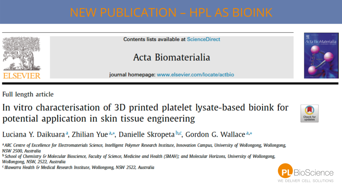
“In vitro characterisation of 3D printed Platelet Lysate-based bioink for potential application in skin tissue engineering”
In a lately published study Daikuara et al. developed an extrusion-based bio-ink consisting of Human Platelet Lysate (PL) and gelatin methacryloyl (GelMA). The bio-ink should be used for the 3D bioprinting of a multifunctional dermal equivalent. For this skin tissue engineering approach human dermal fibroblasts were used.
The combination of a bio-ink with HPL, GelMA and dermal fibroblasts resulted in a stable and functional 3D structure that mimics the extra cellular matrix (ECM). Furthermore, the bioprinted structure protected growth factors (GF) from degradation and enabled spatial-temporal GF delivery for two weeks. (1)
Good cell attachment and improved proliferation
The results of dermal fibroblasts bioprinted with a bio-ink containing HPL and GelMA showed:
- A high cell viability
- Good cell attachement
- Improved proliferation of dermal fibroblasts
- Improved rheological behavior of the bioink
- Upregulated ECM synthesis and deposition by dermal fibroblasts
- Positive impact in the synthesis of ECM components by dermal fibroblasts
The relevance for regenerative medicine
Each year about 8.2 million patients with wounds are treated. Infections and other side effects make wounds are a serious cause for morbidity and mortality.
Wound-related complications and their treatment results in high costs, about 32 billion US Dollar. (1)
The approach to treat wounds with growth factors is very promising. Growth factors (GF) are released by platelets that are initiating the process of haemostastis when we have a wound.
Increasing attention for Human Platelet Lysate in regenerative medicine
Human Platelet Lysate is a rich source for growth factors which are involved in healing and the regeneration of tissues like bone, cardiac tissue, fat or skin.
What is 3D bioprinting?
Using digital files to manufacture biomedical parts of the body. They should mimic the natural tissue.Therefore, layers of cells or materials are printed on each other. These cells or biomaterials are called bio-inks.
A bio-ink can be a mixture of polymers, cells or biomaterials
The rheological behavior, biochemical properties and the biocompatibility of these bio-inks is extremely important.
In general these steps needs to be taken to perform bioprinting:
- Producing a digital file out of CT or MRI data
- Using the bio-ink and the data to create a biomedical structure
Ensure an ideal mechanical and chemical surrounding for cell growth. (2,3)
Why do we want to print organs? What are the advantages?
Organ failure is one of the main causes of mortality all over the world. Organ failure treatments require donated organs for transplantation, a procedure that is related to several problematics: (3)
- Increasing demand for transplanting organs resulting in shortage of supply
- Risk of organ rejection after the transplantation
Therefore, it would be necessary to rise the availability of organs and minimize the risk of rejection.
The advantages of 3D bioprinting are:
- controlled distribution of biomaterials
- controlled distribution of biological molecules
- and allow living cells to form complex 3D structures
Which organs can already be 3D printed?
Until now: no complete functional organs can be bio-printed. The vascular network and complex tissue structure remain to be the bottle neck. (2,3)
But these tissues can already be printed:
- Skin tissue
- Cardiac tissue
- And corneal tissue
3D bioprinting has also an impact on (2,3):
- Automated drug screening
- Controlled cell transplantation
- Metabolism models
- Pathological mechanism analysis
- Cryopreservation of organs/tissues
Get to know more about us and our Human Platelet Lysates: More information
Sources:
(1) https://www.sciencedirect.com/science/article/abs/pii/S1742706121000489?via%3Dihub
(2) https://www.ncbi.nlm.nih.gov/pmc/articles/PMC6952999/
(3) https://www.news-medical.net/health/What-is-Bioprinting.aspx